Field Guide to Histology
Print ISBN: 978-1-68135-481-1
Help flip the histological components of your laboratory. When students use this “field guide” to identify unknown samples, they are quickly able to differentiate sources based on similar traits and appearance. When used in conjunction with their lab manual or textbook, the names of the differentiating characters they used are then learned, lessening confusion and improving mastery. Students learn the way they learn, not the way the instructor learns.
Outstanding Features:
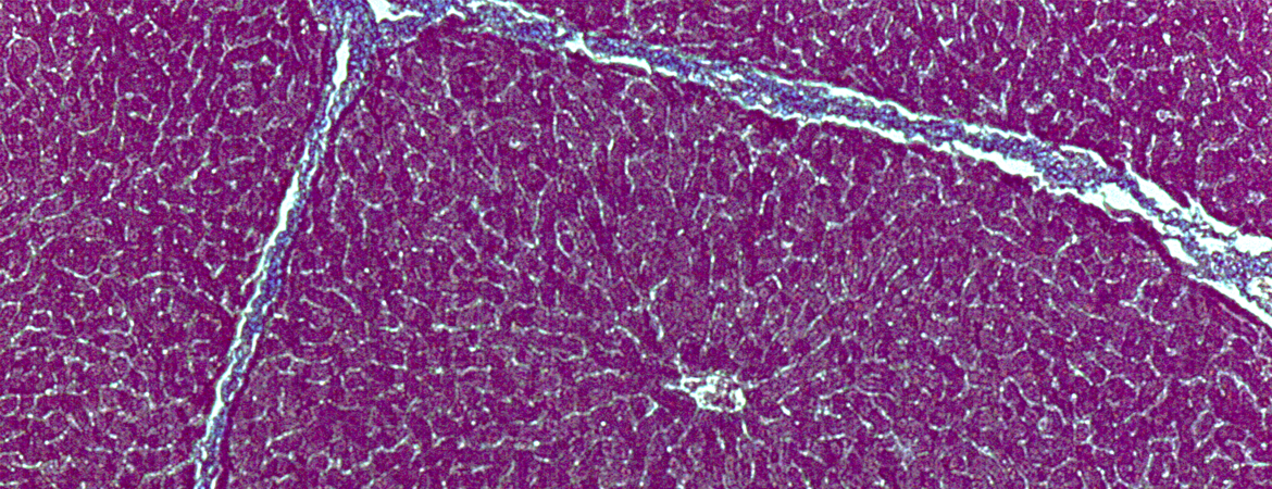
- Full-color images of cells, tissues, and organs common to the Anatomy and Physiology laboratory.
- Allows students to quickly reference topics such as epithelial, nervous, connective tissues, muscles, and organs.
- Images presented at magnifications typically used in the laboratory. Many images presented at multiple magnifications. This helps guide students to know which power they should be using in class.
- Convenient and practical resource for students.
Table of Contents
- Epithelial Tissues
- Connective Tissues
- Muslce
- Nervous Tissue
- Organs
- Special Cells or Structures of Interest
A Look Inside
About the Author
John Cummings has been an instructional faculty member at Clemson University since 1989. He runs an integrated, two-semester Human Anatomy and Physiology course sequence, with enrollments right at 500 students. He was recognized with the College of Agriculture Forestry and Life Science Outstanding Teaching Award and the Clemson University Alumni Master Teacher Award. He completed both his undergraduate and graduate work at Bowling Green State University in Ohio.